Breast-conserving surgery (lumpectomy)
This operation consists of removing only the tumour and a margin of healthy tissue around it. It is generally followed by radiotherapy.

Breast cancer is the most common cancer in women. 1 woman in 8 is at risk of developing breast cancer during her lifetime. Early detection and therapeutic advances now make high cure rates possible.
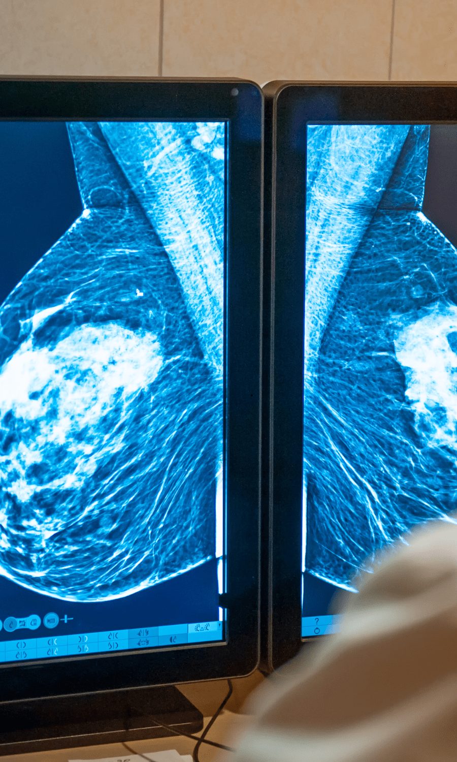
Breast cancer develops from the cells of the mammary gland. There are different types of breast cancer, each with its own characteristics that will guide the choice of treatments:
Ductal carcinoma in situ (DCIS) This is an early form of the cancer, in which the cancer cells are confined to the milk ducts (which carry milk to the nipple). At this stage, the cancer cells have not spread into the surrounding tissue. Early diagnosis of this form generally allows an excellent prognosis.
Invasive ductal carcinoma This is the most common form, accounting for around 80% of breast cancers. The cancer cells have crossed the wall of the ducts and begun to invade the surrounding breast tissue. Without treatment, these cells can spread to the lymph nodes or other parts of the body.
Lobular carcinoma It develops in the lobules, the glands that produce breast milk. It exists in two forms:
Several factors can increase the risk of developing breast cancer:

Breast cancer treatment is personalised according to several criteria:
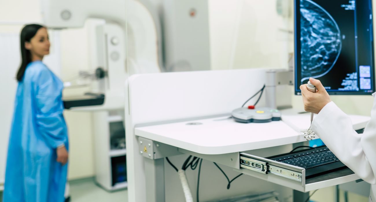
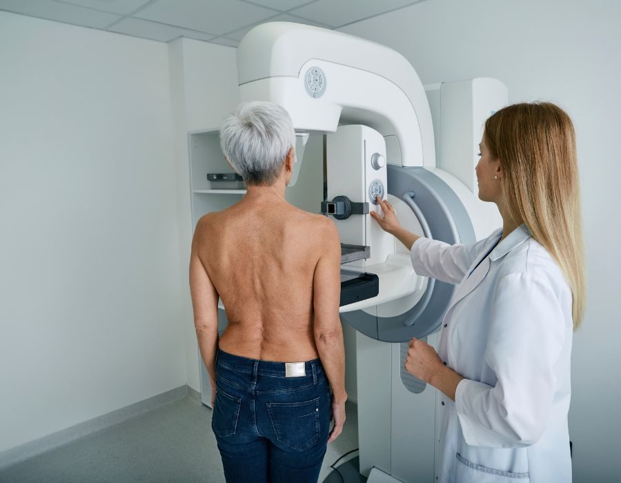
Early screening is crucial because it makes it possible to detect the cancer at a stage where the chances of cure are highest. In France, a national organised screening programme offers a free mammogram every 2 years to women aged between 50 and 74.
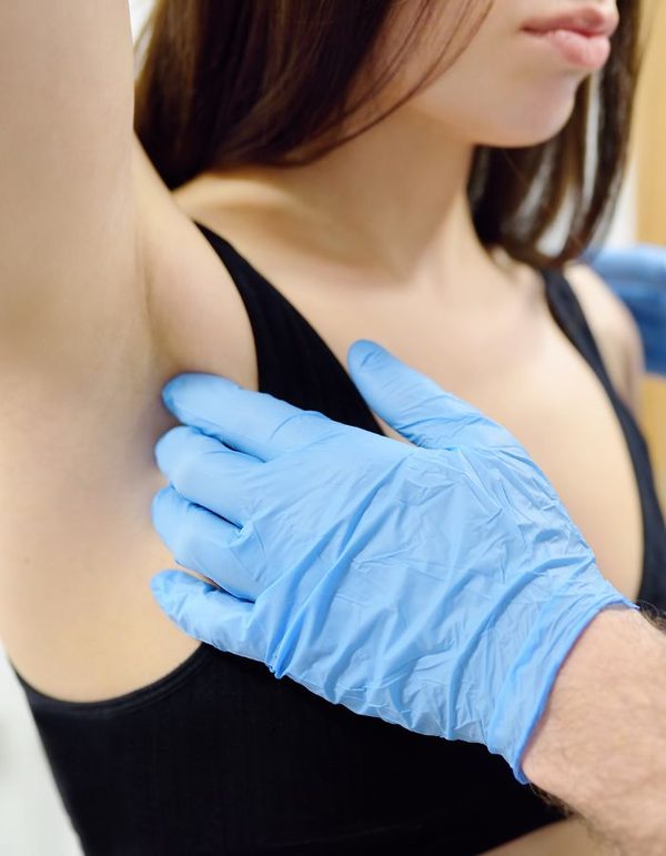
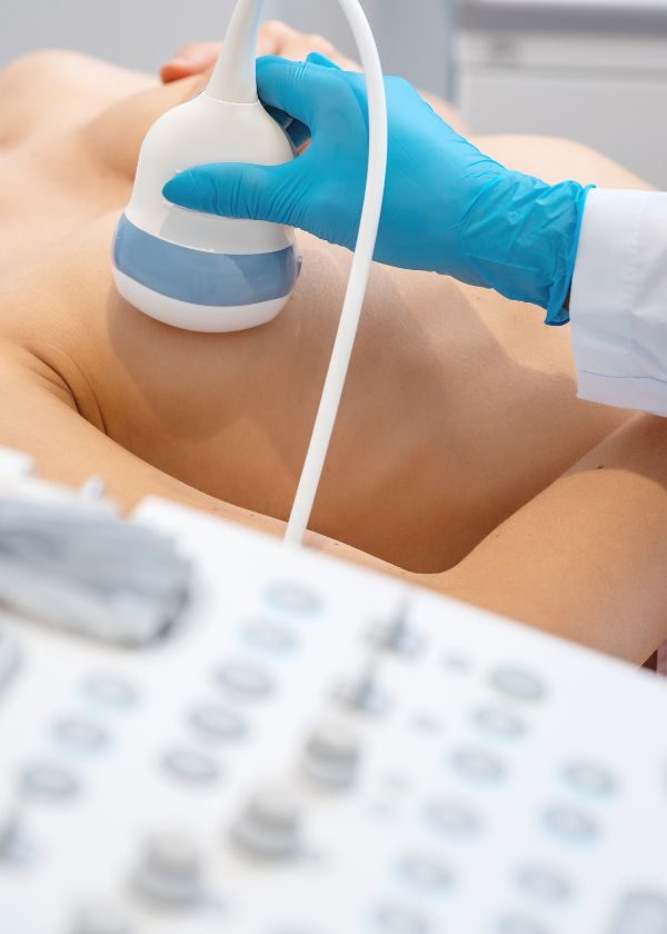
Breast cancer detection relies on several examinations:
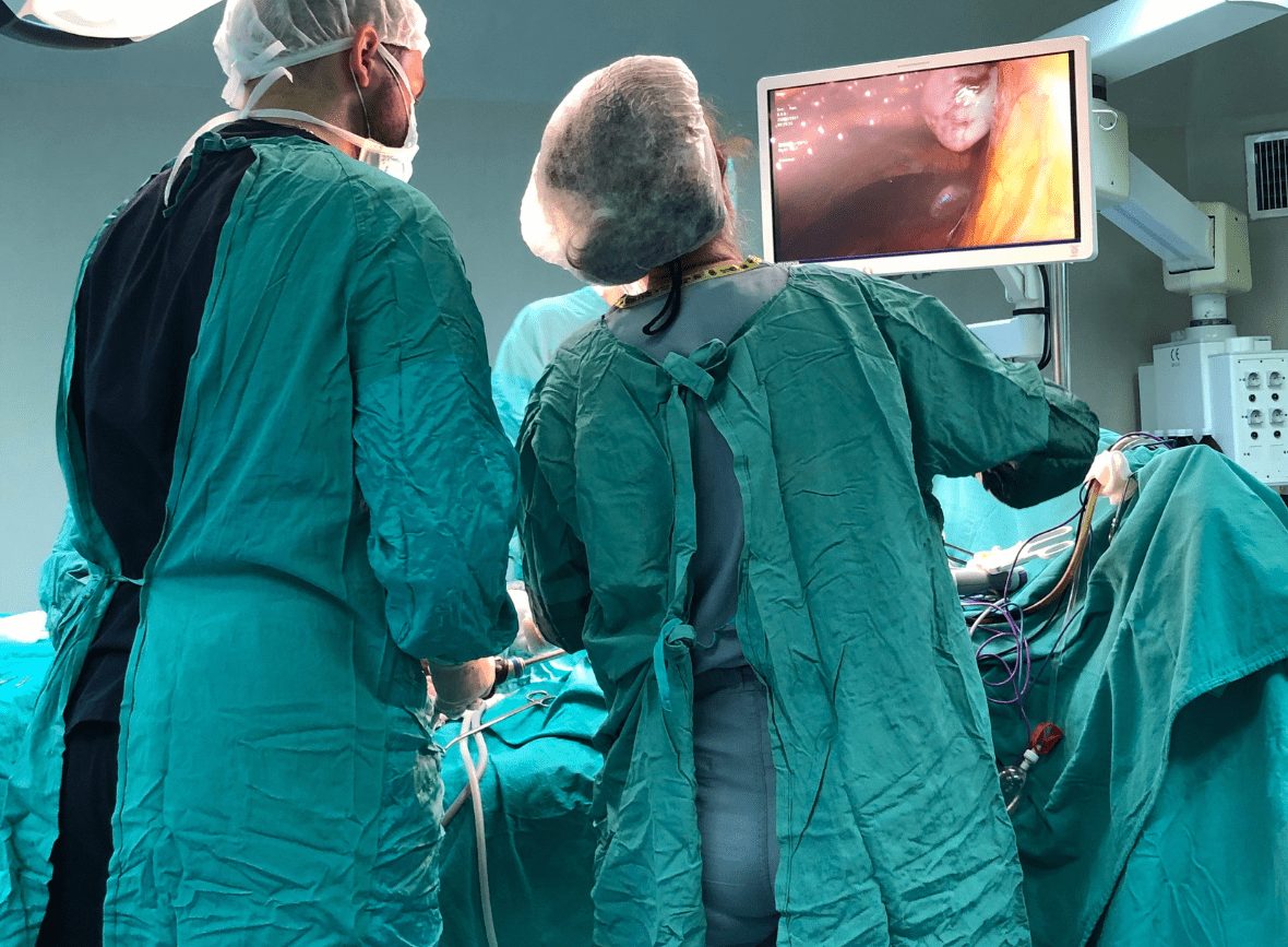
Surgery is often the first treatment offered. 2 types of operation are possible:
This operation consists of removing only the tumour and a margin of healthy tissue around it. It is generally followed by radiotherapy.
In some cases, the whole breast has to be removed. Breast reconstruction can then be offered, either immediately or at a later stage. The surgeon also examines the lymph nodes in the armpit, either using the sentinel lymph node technique or through a more extensive axillary dissection.
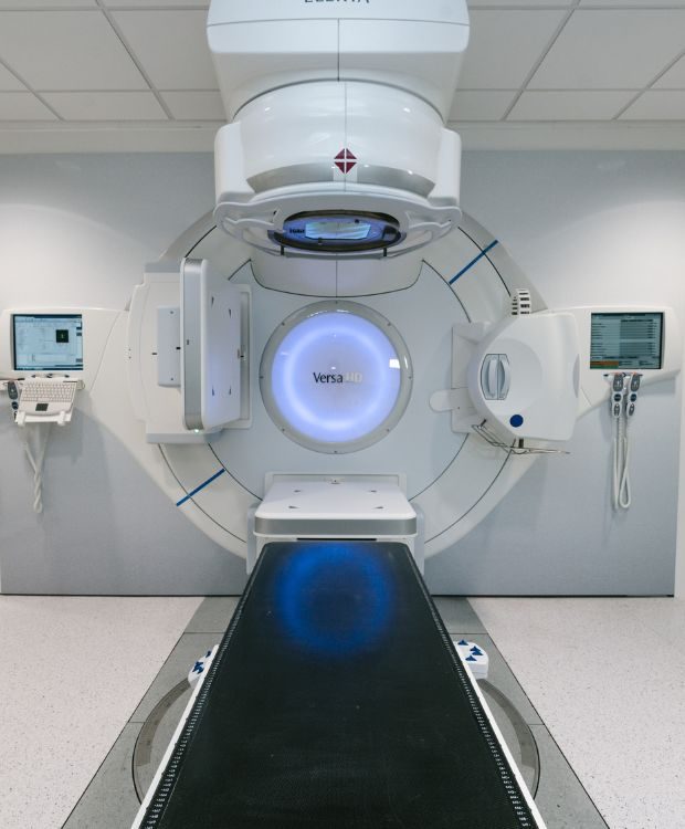
Radiotherapy: This treatment uses radiation to destroy residual cancer cells. It is systematic after breast-conserving surgery and sometimes necessary after a mastectomy.
Chemotherapy: It can be prescribed before surgery (neoadjuvant chemotherapy) or after it (adjuvant chemotherapy). Its aim is to destroy cancer cells that may have spread.
Hormone therapy: For hormone-sensitive cancers, this treatment blocks the action of the hormones that stimulate the growth of cancer cells. It is generally prescribed for several years.
Targeted therapies: These more recent treatments specifically target certain characteristics of cancer cells.
Organised screening is recommended between the ages of 50 and 74, with a mammogram every two years. However, in the event of a family history or risk factors, personalised follow-up can be set up earlier. Consult your doctor to define the schedule best suited to your situation.
The examination takes around 15 minutes. Each breast is compressed between two plates to obtain good-quality images. Although the compression can be uncomfortable, it is brief and essential for an accurate diagnosis.
The total duration varies according to the type of cancer and the treatments required. Surgery requires a few weeks of recovery. Radiotherapy generally lasts 5 to 6 weeks. Chemotherapy can extend over 4 to 6 months. Hormone therapy, if necessary, is prescribed for several years.
This depends on the type of treatment, your general health and your occupation. Some patients keep working, others prefer to take sick leave. Adjustments to your position or therapeutic part-time work can be considered.
No, it is a personal choice. Reconstruction can be immediate or delayed, depending on your wishes and medical recommendations. Some women choose not to have reconstruction.
Some treatments, particularly chemotherapy, can affect fertility. If you are planning a pregnancy, discuss it with your medical team before the treatments begin. Fertility preservation options can be offered.
The effects vary according to the treatments: fatigue, nausea, digestive problems, hot flushes, joint pain… Your medical team can help you prevent and relieve them. Most are temporary and disappear after the treatments.
The risk exists, but decreases over time. Regular follow-up makes it possible to detect and treat any recurrence quickly. Keeping to the surveillance schedule is essential.
Adapted physical activity is recommended: it improves tolerance of the treatments and reduces fatigue. Talk to your medical team, who can direct you towards suitable programmes.
Many associations offer valuable support and guidance throughout your journey. Click here to discover a selection of patient associations that can accompany you.