Skip to content
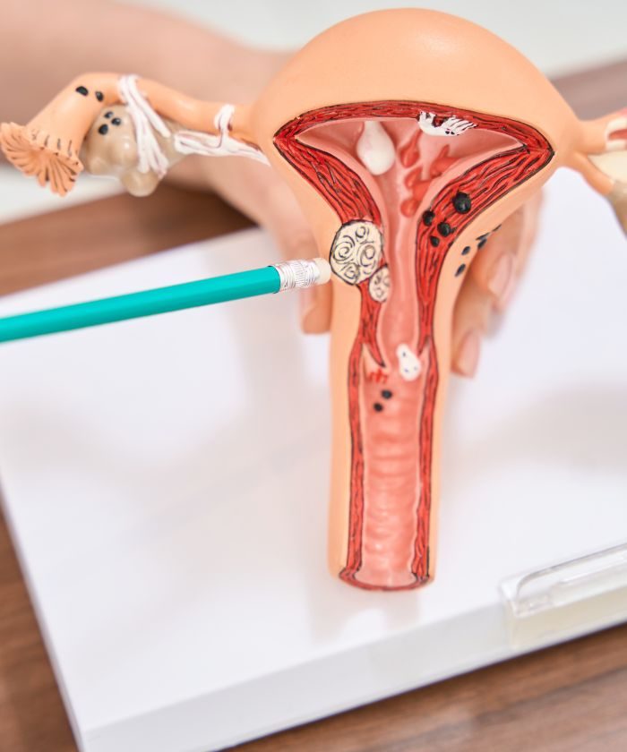
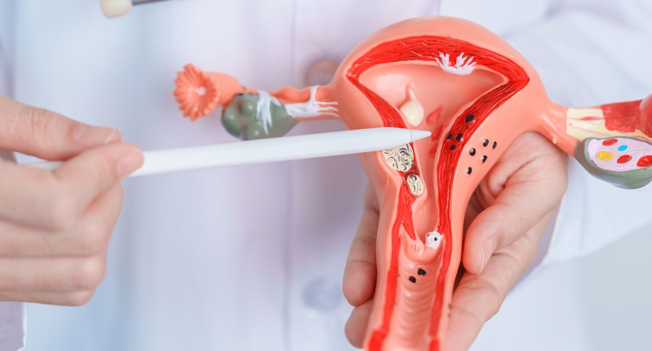
Doctor pointing to the endometrium on an anatomical model of the uterus

Endometrial cancer

Endometrial cancer, also called cancer of the uterine body, develops in the lining of the inside of the uterus. It is the most common gynaecological cancer after breast cancer, affecting mainly women after the menopause.

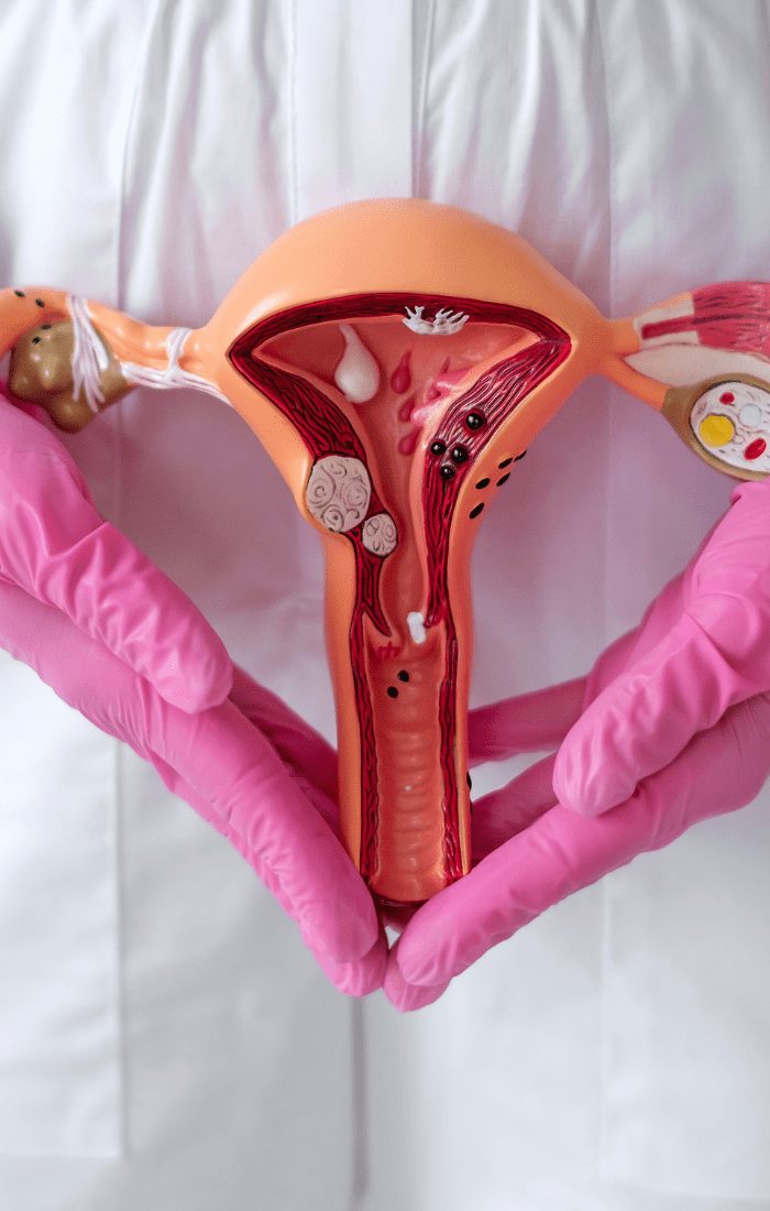
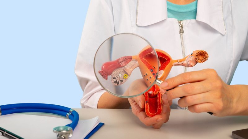
Gloved hands holding an anatomical model of the uterus

Understanding endometrial cancer

The majority of endometrial cancers develop from the glandular cells of the uterine lining. Several types are distinguished:

Endometrioid adenocarcinoma This is the most common form, accounting for around 80% of cases. It develops from the glandular cells of the endometrium. These cancers are generally diagnosed at an early stage, because they cause bleeding quickly, which often allows prompt treatment with a good prognosis.

Serous carcinoma Rarer but more aggressive, it accounts for around 10% of endometrial cancers. It requires specific management because it tends to spread more quickly than the endometrioid form.

Clear cell carcinoma This form is also uncommon (around 5% of cases). Like serous carcinoma, it is considered more aggressive and requires adapted management.

Mixed forms Some cancers may show the characteristics of several types. The treatment is then adapted according to the components present.

This classification is important, because it guides treatment decisions. Each type of cancer may require a different approach in terms of surgery and complementary treatments.

What are the risk factors?

Several factors can increase the risk:

  • Age: more common after the menopause
  • Obesity
  • Diabetes
  • High blood pressure
  • No pregnancy
  • Early puberty or late menopause
  • Taking tamoxifen (a breast cancer treatment)
  • Certain family histories (Lynch syndrome)
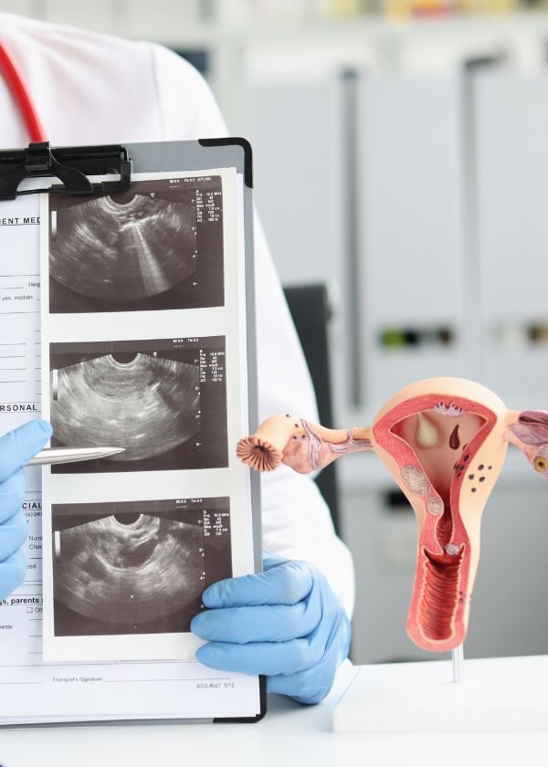
Doctor holding an ultrasound image and a model of the uterus Patient receiving an infusion treatment
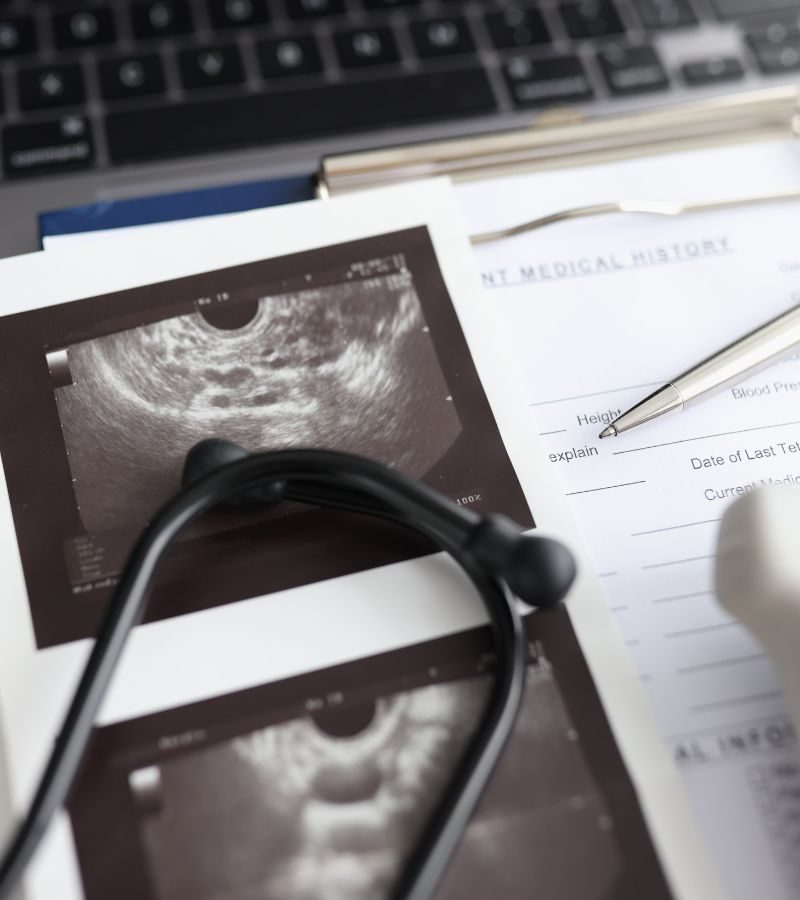
Stethoscope resting on ultrasound images

Treatment approach

Treatment is personalised according to:

  • The type and stage of the cancer
  • The patient's age
  • Her general health
  • Any wish for a pregnancy

Detection and diagnosis

The main warning sign is vaginal bleeding:

  • After the menopause: any bleeding is abnormal and should prompt a consultation
  • Before the menopause: irregular or abnormally heavy bleeding
  • Unusual vaginal discharge
Anatomical model of the uterus on a desk

Diagnostic examinations

The diagnosis relies on several examinations:

  • Gynaecological examination
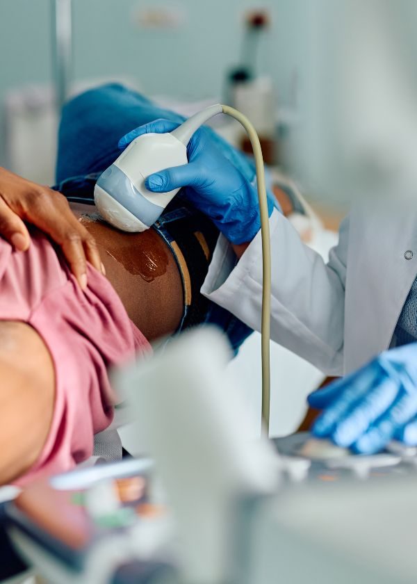
  • Pelvic and transvaginal ultrasound
  • Hysteroscopy: allows the inside of the uterus to be visualised
  • Endometrial biopsy
  • Pelvic MRI
  • Chest, abdominal and pelvic CT scan
  • PET scan in some cases
Laparoscopy instruments on a surgical drape

Surgical procedures

Total hysterectomy: This is the complete removal of the uterus and cervix. This operation can be performed by different routes depending on your situation:

  • Minimally invasive route (laparoscopy): small incisions in the abdomen
  • Vaginal route: no scar on the abdomen
  • Laparotomy: a conventional abdominal incision, reserved for certain specific cases The choice of technique depends on several factors that your surgeon will assess with you.

Removal of the ovaries and fallopian tubes: Called bilateral adnexectomy, this operation is generally performed at the same time as the hysterectomy. It is recommended because the ovaries can be a site of cancer spread, and their presence can in some cases stimulate the growth of cancer cells. In women who have not reached the menopause, this operation will cause an immediate menopause that will require specific support.

Pelvic lymph node dissection: This step consists of removing the lymph nodes of the pelvis for analysis. It is an important procedure that checks whether the cancer has spread to the lymph nodes and determines the precise stage of the disease, so that complementary treatments can be adapted if necessary. The less invasive sentinel lymph node technique can sometimes be offered in certain cases.

Complementary treatments

Radiotherapy

Radiotherapy uses radiation to destroy cancer cells that may persist after surgery. It can be given in two ways: either externally, where the radiation passes through the skin to reach the pelvis, or by brachytherapy, where a radioactive source is placed directly in the vagina for a more targeted treatment.

Chemotherapy

Chemotherapy is a treatment that circulates throughout the body via the bloodstream to destroy cancer cells. It is offered in certain specific situations: when the cancer is at an advanced stage, when it is growing rapidly, or if there are significant risk factors for recurrence.

Hormone therapy

Hormone therapy can be offered in certain cases of endometrial cancer that are sensitive to hormones (positive hormone receptors). It works by blocking the action of the hormones that could stimulate the growth of cancer cells. This treatment is particularly used in the event of recurrence, for advanced cancers, or in some young women wishing to preserve their fertility when the situation allows it.

Injection given in a patient's arm

Frequently asked questions

How is a hysteroscopy performed?

It is a quick examination that allows the inside of the uterus to be examined using a small camera. It is performed in the consulting room or under light anaesthesia.

Is radiotherapy painful?

Radiotherapy is not painful during the sessions, but it can cause side effects that will be monitored and managed.

Will I be able to have children after the treatments?

This is an important question, particularly for young women. In the majority of cases, the standard treatment for endometrial cancer includes removal of the uterus and ovaries, making a future pregnancy impossible. However, for some young patients with a very early cancer, it is sometimes possible to discuss a treatment that preserves fertility. This decision is taken after an in-depth discussion with the medical team, taking into account your personal situation and the characteristics of your cancer. Do not hesitate to raise your wishes for a pregnancy with your doctor from the very start of your care.

What are the possible side effects?

They vary according to the treatments: fatigue, digestive problems, hot flushes… The medical team supports you in managing them as well as possible.

What does follow-up after the treatments involve?

A personalised surveillance schedule is drawn up. Follow-up after treatment includes:

- Regular clinical examinations

- Pelvic ultrasounds

- Blood tests

- Other imaging examinations depending on the case

What are the possible side effects of the treatments?

The effects vary according to the treatments: fatigue, nausea, digestive problems, risk of infection… The medical team supports you in managing them as well as possible.

Can I resume physical activity after surgery?

Resuming physical activity is gradual and encouraged. Your medical team will guide you on the appropriate pace.

Does the treatment affect intimate life?

Changes to intimate life are possible. Do not hesitate to discuss them with your medical team, who can advise and guide you.

Are all the treatments carried out in hospital?

Some treatments require hospitalisation, others can be carried out as outpatient care. The programme is adapted to each situation.

Are there support groups?

Many associations offer valuable support and guidance throughout your journey. Click here to discover a selection of patient associations that can accompany you.