Skip to content
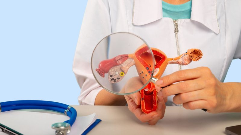
Gloved hands presenting an anatomical model of the uterus

Uterine cancer

Uterine cancer, or cancer of the uterine body, develops in the body of the uterus. It is the fourth most common cancer in women, affecting mainly women after the menopause.

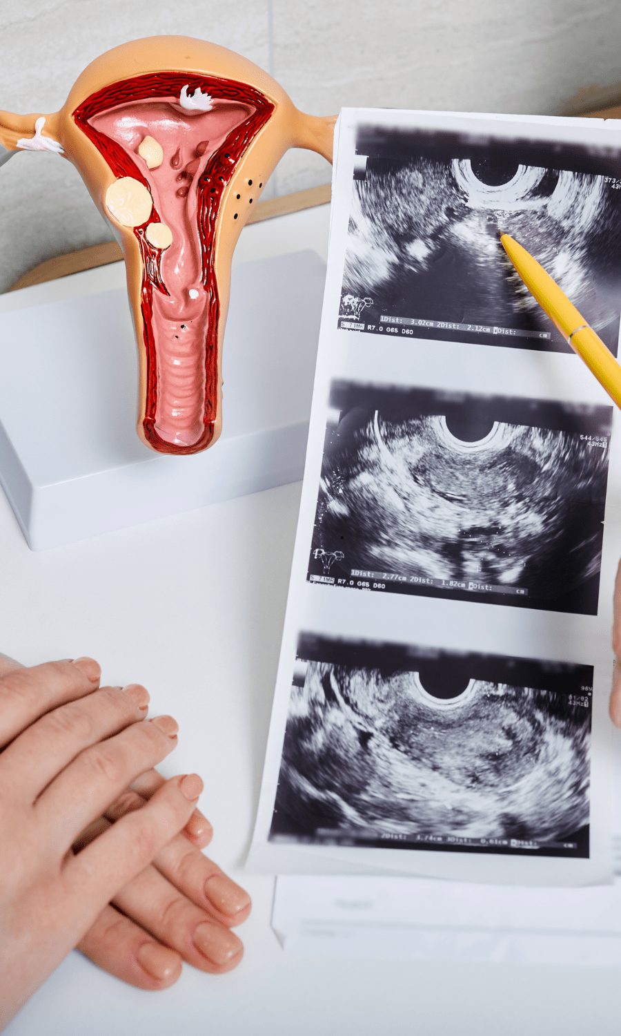
Model of the uterus and ultrasound images on a desk

Understanding uterine cancer

Uterine sarcomasThese cancers develop in the muscular part of the uterus. Imagine the uterus as a pouch whose wall is made of a thick muscle. It is in this muscle that sarcomas develop. Several types are distinguished:

  • Leiomyosarcoma: this is the most common form. It develops directly in the muscle fibres of the uterus, a little like a fibroid, but with cancer cells.Endometrial stromal sarcoma: rarer, it affects the tissue that supports and nourishes the uterine lining (the endometrium). It is as if the "skeleton" that holds the lining were affected.Undifferentiated sarcomas: in these forms, the cancer cells have changed so much that it is difficult to recognise their precise origin. These forms are more aggressive and require rapid treatment.

What are the risk factors?

Several factors can increase the risk:

  • Age: more common after 60
  • Obesity
  • High blood pressure
  • Diabetes
  • A history of pelvic radiotherapy
  • Certain hormone treatments
  • A family history of cancers
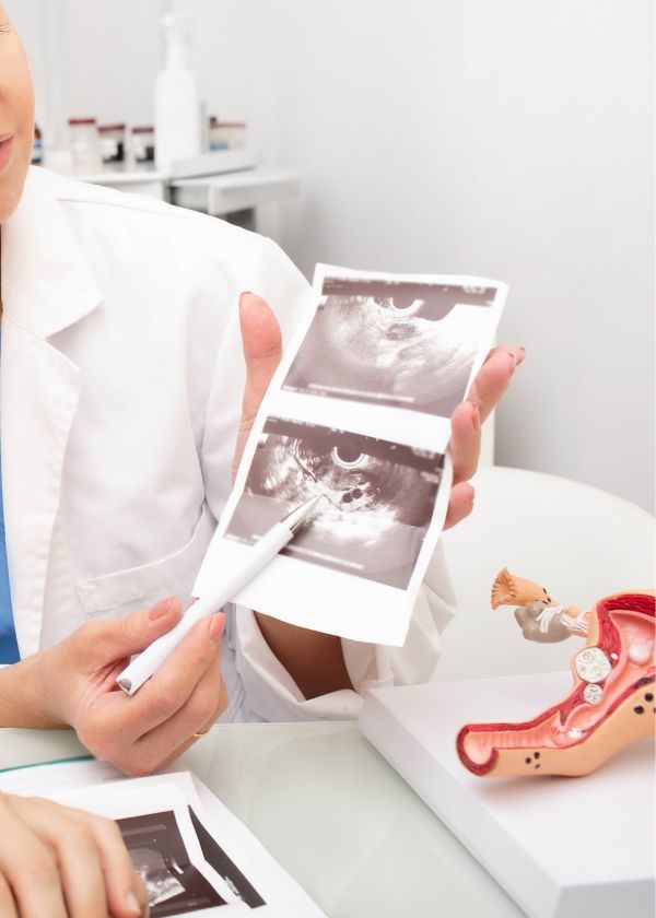
Model of the uterus and ultrasound image on a consulting desk Doctor showing ultrasound images
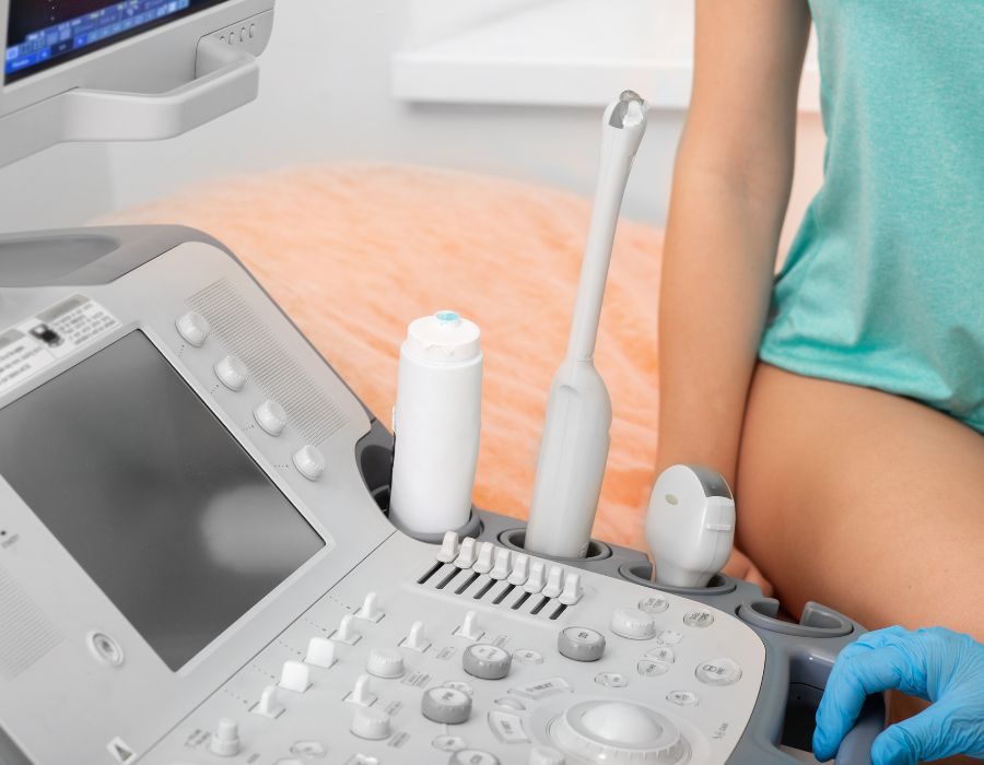
Ultrasound scanner and probe in an examination room

Treatment approach

Treatment is personalised according to:

  • The type and stage of the cancer
  • The size of the tumour
  • The patient's age and general health

Screening and diagnosis

Uterine cancer manifests itself mainly as abnormal vaginal bleeding. After the menopause, any loss of blood, however small, should be considered abnormal and requires a prompt consultation. Before the menopause, be attentive to changes in your periods: bleeding that becomes irregular or particularly heavy should alert you. Other signs may also appear, such as pain in the lower abdomen (pelvic pain) or unusual vaginal discharge.

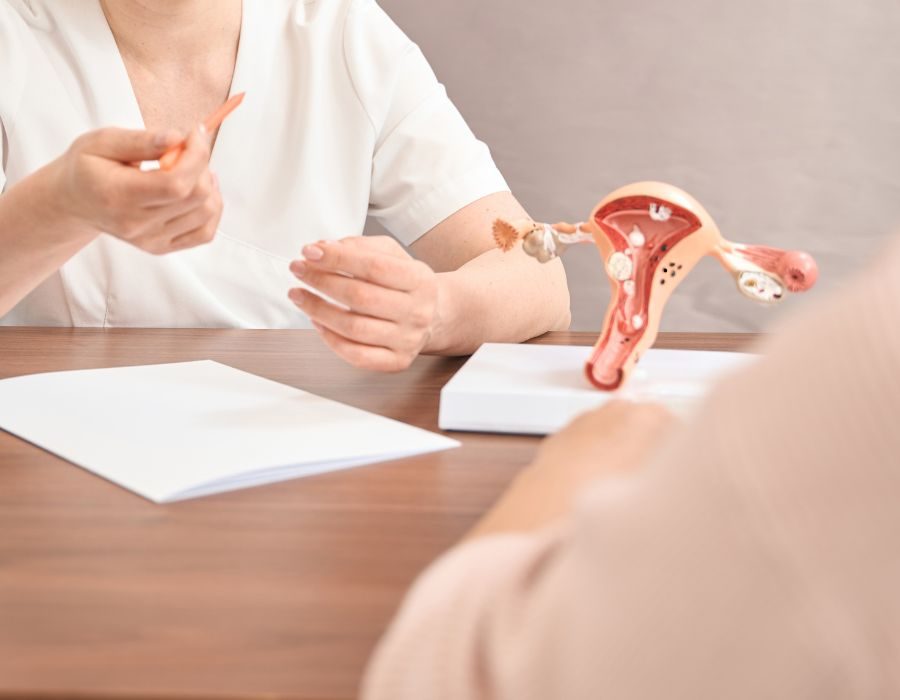
Consultation: model of the uterus on the desk

Diagnostic examinations

The diagnosis relies on several examinations:

  • Clinical gynaecological examination
  • Pelvic and transvaginal ultrasound
  • Pelvic MRI
  • Uterine biopsy
  • Chest, abdominal and pelvic CT scan
  • PET scan in some cases

Surgical procedures

Complete removal of the uterus and cervix

The whole uterus is removed, including the cervix, to make sure that no cancer cells remain. This removal can be performed by different routes depending on your situation:
• By laparoscopy: a minimally invasive technique with small incisions in the abdomen
• By conventional abdominal surgery: necessary in certain specific cases

Removal of the ovaries and fallopian tubes

The ovaries and fallopian tubes are also removed, because they can be a site of cancer spread. In women who have not reached the menopause, this removal will cause an immediate menopause that will be medically supported.

Removal of the lymph nodes

The lymph nodes of the pelvis are removed and analysed to check whether the cancer has spread there. This information is crucial for adapting the rest of the treatment.

Surgical instruments handled by gloved hands
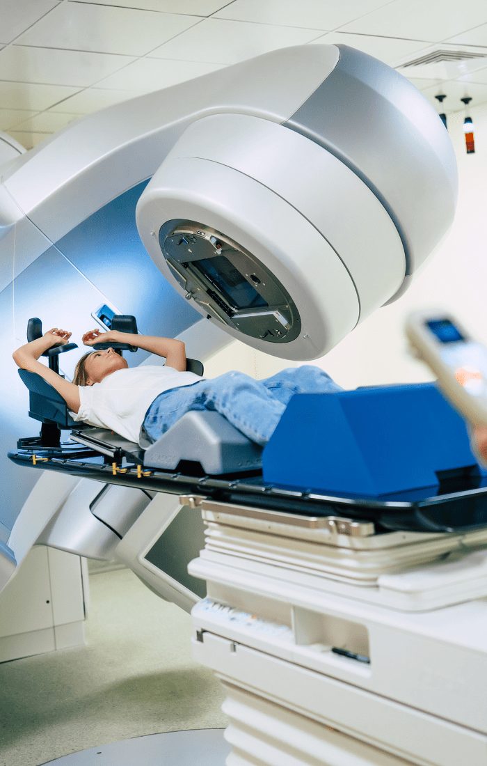
Patient positioned on a radiotherapy machine

Complementary treatments

Depending on the characteristics of your cancer, other treatments may be necessary:

Radiotherapy: It uses radiation to destroy cancer cells that may have remained after surgery. It can be given in two ways:

  • From the outside (external radiotherapy): the radiation passes through the skin to reach the area to be treated
  • From the inside (brachytherapy): a radioactive source is placed in direct contact with the area to be treated

Chemotherapy: This drug treatment circulates throughout the body to destroy cancer cells. It is offered in certain situations:

  • When the cancer is aggressive
  • If there is a significant risk of recurrence
  • If cancer cells have been found in the lymph nodes

The choice and order of these treatments are always discussed at a multidisciplinary team meeting to offer you the strategy best suited to your situation.

Frequently asked questions

What are the first signs of uterine cancer?

Abnormal vaginal bleeding, particularly after the menopause, is the most common sign. Any loss of blood after the menopause should prompt a rapid consultation.

What are the side effects of a hysterectomy?

In women who have not reached the menopause, removal of the ovaries causes an immediate menopause. The operation has no impact on intimate life after the healing period.

Is radiotherapy painful?

Radiotherapy is not painful during the sessions, but it can cause side effects (fatigue, local irritation) that will be monitored and managed by the medical team.

What impact on my intimate life?

After the healing period (around 6-8 weeks), you will be able to resume an intimate life. The operation does not alter sensation or pleasure, but there may be a period of adjustment. Do not hesitate to discuss this with your medical team.

Will there be effects on my mood and my energy?

It is normal to feel fatigue and mood changes during and after the treatments. Psychological support can be beneficial in getting through this period. Professionals are there to accompany you.

What are the effects of surgical menopause?

If you had not reached the menopause, removal of the ovaries will cause menopausal symptoms (hot flushes, mood changes, etc.). Hormone replacement therapy can be discussed with your doctor depending on your situation.

How do I know whether the treatment is working?

Effectiveness is assessed through regular examinations during and after the treatments. Your medical team will explain the results of each examination to you and adapt the follow-up accordingly.

Are there support groups?

Many associations offer valuable support and guidance throughout your journey. Click here to discover a selection of patient associations that can accompany you.